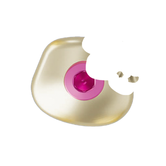
Restauración posterior cementada de una sola pieza de la mandíbula mediante un protocolo iPhysio
This case study illustrates all the steps involved in creating a prosthetic restoration using the iphysio protocol on ETK Naturactis implants.
The patient was 66 years old, in good general health, a non-smoker and in need of implant-prosthetic rehabilitation of tooth 46.
The treatment plan was as follows:
- Tooth 46 extracted in January 2018
- Placement of a 10 mm Naturactis Ø 5 implant
- Given the implant's good primary stability, an iphysio® Profile Designer, shape B, height 1 mm, was placed in immediate loading.
- The optical impression is taken two months later.
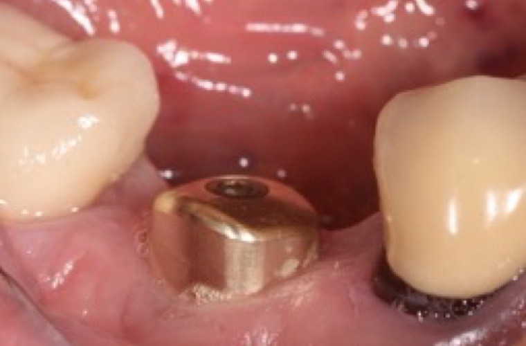
1. iPhysio® Profile Designer (shape C - height 1) screwed in position 46
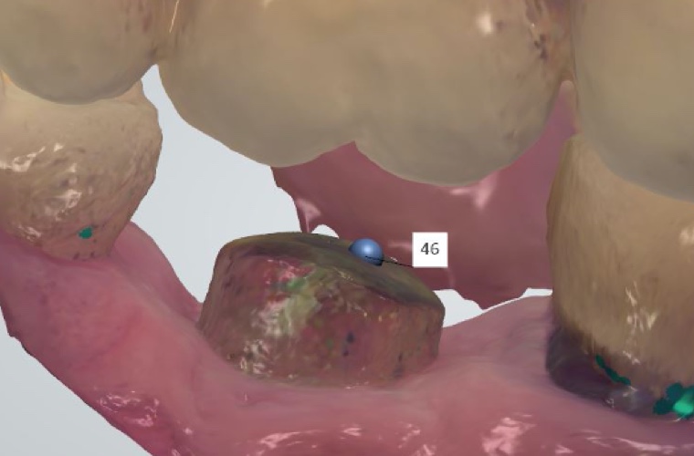
2. Profile Designer iPhysio® CT scan with Trios 3Shape intraoral scanner
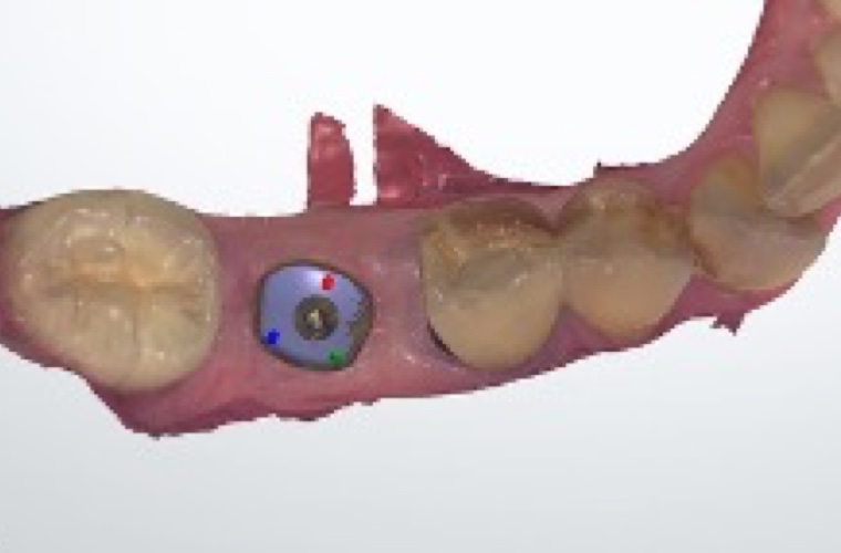
3. Positioning of the iPhysio® replica from the library using a point on the iPhysio® CT scan.
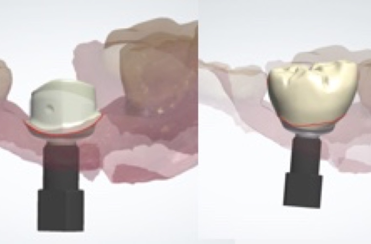
4. Customized abutment design retaining the gingival anatomical profile of the iPhysio® Profile Designer and crown design
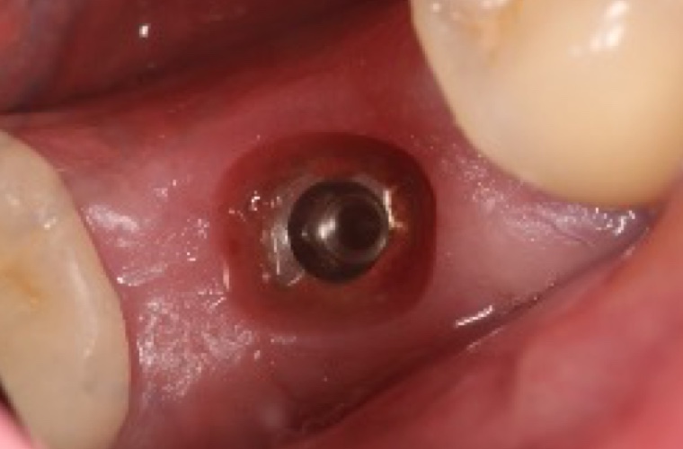
5. iPhysio® Profile Designer removed. The anatomical profile of the prosthetic cradle can be seen.
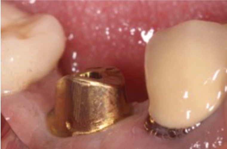
6. Screwing a custom abutment into the mouth
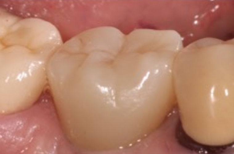
7. Final result after crown cementation
